

PROJECT DETAILS
- Hospital Name Prabhu Netralaya
- Doctor Dr. Ankit Gupta
- Surgery Retinal Laser Treatment
- Date August 16, 2024
- Diseases Retinal Laser Treatment
- Categories General Patient
- Location Rudrapur
LASER
LASER stands for Light Amplification by Stimulated Emission of Radiation. It is a high energy beam of light which is used to treat the eyes in various ways. LASERs used in the OPD (Out-patient department) are mainly used for the management of retinal and anterior segment conditions.
Retinal LASER:
These are done usually to the swelling in the central part of the retina (Macular oedema), for areas of ischaemia in retina especially when abnormal new vessels are formed or to weak areas in retina (holes, tears).
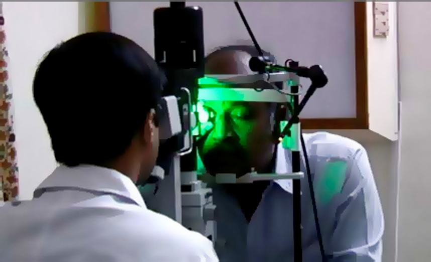
Procedure and preparation:
The eyes will need to be dilated prior to the treatment. LASER can be delivered either by indirect ophthalmoscope (LIO) or by slit lamp. LIO is usually preferred if the area to be treated is in the far periphery or when the media is very hazy due to cataract or vitreous haemorrhage. A contact lens needs to be applied on the eye before slit lamp delivery of LASER. Hence, anaesthetic drops are applied before commencing slit lamp delivery of LASER. The procedure usually lasts for about 5-15 minutes.
Red LASER (810 nm):
Due to its larger wavelength, the LASER penetrates deeper tissues. It also penetrates better in eyes with retinal haemorrhage. It is usually used for treating the abnormal retina in babies with Retinopathy of prematurity (ROP)
Green LASER (532nm):
This is the conventional form of retinal laser and gives the best result in a clear media. This LASER can be delivered by LIO or by slit lamp. Our hospital has facilities to deliver this LASER through slit lamp as a single spot or multiple spot LASER.
Yellow LASER (577nm):
Adding to our unrelenting efforts to be the best in Retina and Eye care, we introduced in 2014, for the first time in AP & Telangana, the Yellow LASER, for the treatment of multitude of macular diseases like diabetic macular oedema, CSCR and ARMD. This makes us one of the handful of Eye hospitals across the country to possess such remarkable and state-of-the-art equipment. The Yellow LASER also heralds a new beginning in the area of Diabetic Retinopathy, as it has been proven to be extremely effective, besides being safer than the existing technology. This LASER has an ability to penetrate media opacities like cataract and vitreous haemorrhage as it has minimal light scattering property thereby providing more accurate treatment. Unlike conventional green LASER, the Yellow LASER has more consistent effect on patients with irregular or light fundus pigmentation as it is less dependent on melanin in retinal pigment epithelium (RPE) and more dependent on haemoglobin on choriocapillaris. It is minimally absorbed by macular pigments (xanthophylls) and well absorbed by oxygenated haemoglobin. It is also the ideal wavelength for micro pulse sub threshold laser therapy used for diabetic macular oedema, branch retinal vein occlusions and central serous retinopathy. With this mode, exposures as low as 10 milliseconds are possible, hence enabling LASER treatment with no visible endpoint or tissue damage. Due to these enhanced safety features it is the treatment of choice for macular LASERs. Our hospital has facilities to deliver this LASER through slit lamp as a single spot or multiple spot LASER.
Disadvantages of conventional LASER:
More pain as exposure time cannot be less than 100 ms
Worsening of macular oedema. Multiple sessions of conventional LASER are advocated in order to overcome this disadvantage Expansion of spot size resulting in scarring and RPE atrophy.
Visual field defects and reduced dark adaptation
The above disadvantages result in a poorer patient experience thereby adversely affecting their compliance to the therapy
Multiple spot LASER:
We are also the first in AP and Telangan to acquire the latest Multispot Pattern LASER photocoagulator. Here multiple spots of LASER can be delivered to the retina in a pre-fixed pattern. The exposure time is drastically reduced resulting in a decreased sensation of pain by a factor of up to 4.5 times lesser than conventional LASER. This results in faster treatment sessions (reduced by a factor of 10) resulting in lesser discomfort to the patient. The spots are uniform and well defined resulting in preservation of the sensitive inner retinal layers. This reduces the incidence of poorer night vision or reduced visual field. There is no expansion of the spots with no RPE atrophy or scarring. So there is minimal collateral damage. There is also minimal inflammation; therefore, reducing the incidence of LASER induced macular oedema. Overall it results in an improved patient experience.

Indications for retinal lasers:
– Diabetic Macular oedema
– Proliferative diabetic retinopathy
– Macular oedema due to retinal vein occlusions
– Neovascularisation secondary to retinal vein occlusions
– Peripheral retinal degenerations/holes/tears
– Eales disease and other retinal vasculitis
– Central serous retinopathy
– Retinopathy of prematurity
– Extrafoveal polyps of polypoidal choroidal vasculopathy (PCV)
Rarely
- – Coat’s disease
– Familial exudative vitreo retinopathy
– Retinal Artery Macroaneurysm
– Ocular ischaemic syndrome with secondary neovascularisation
